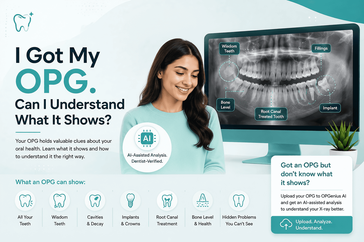
I Got My OPG. Can I Understand What It Shows?
Got an OPG but not sure what you're looking at? Learn how to understand your panoramic dental X-ray, what teeth, wisdom teeth, fillings, implants, bone levels, and other findings may look like.
You finally get your dental X-ray.
You open it on your phone and stare at it for a few seconds.
Teeth. Roots. Shadows. White areas. Dark areas.
And then you think:
“Okay… but what am I actually looking at?”
If you've recently had an OPG, you're probably not alone.
You may be wondering:
What do all these areas mean? Can I spot a cavity? Are my wisdom teeth okay? Is that dark area normal?
The good news is that you can understand the basics of an OPG without being a dentist.
But there's an important distinction:
Understanding what you see is not the same as diagnosing what's wrong.
Let's take a look.
First, what exactly is an OPG?
An OPG, also called a panoramic dental X-ray, is a type of dental imaging that captures a broad view of your:
- Upper and lower teeth
- Upper and lower jaws
- Wisdom teeth
- Tooth roots
- Surrounding bone
- Some surrounding anatomical structures
Unlike a small dental X-ray that focuses on just a few teeth, an OPG provides a panoramic overview of the mouth and jaws in a single image.
That's why dentists may recommend an OPG when they need to see the bigger picture.
So, what can you actually see on an OPG?
Let's make it simple.
1. Your teeth
This might seem obvious, but your dentist can assess all the teeth visible in the image, including their:
- Position
- Shape
- Development
- Roots
- Relationship with neighbouring teeth
You may also be able to spot things like crowding, missing teeth, or teeth that haven't erupted properly.
2. Your wisdom teeth
Look all the way at the back of the image.
See those last teeth?
Those are your wisdom teeth, if you have them.
An OPG can help show whether wisdom teeth are:
✔ Fully erupted
✔ Partially erupted
✔ Impacted
✔ Angled toward another tooth
✔ Developing underneath the gums
And here's something many patients don't realize:
A problematic wisdom tooth doesn't always cause pain.
That's one reason dentists may recommend an OPG when evaluating wisdom teeth.
3. Dark and white areas, what do they mean?
This is probably the part that makes patients curious.
Why are some areas of an OPG white, while others look dark?
In simple terms, different tissues absorb X-rays differently.
Structures that are denser generally appear lighter or whiter, while areas that allow more X-rays to pass through appear darker.
For example, teeth and bone appear relatively light, while air spaces appear darker.
But here's where you need to be careful:
A dark spot doesn't automatically mean you have a cavity.
And a white area doesn't automatically mean something is wrong either.
The appearance of an area on an X-ray needs to be interpreted alongside the location, shape, clinical examination, symptoms, and sometimes other types of dental imaging.
So don't zoom into one mysterious shadow and start Googling:
“Is this serious?”
4. Can you see cavities on an OPG?
Sometimes dental decay may be visible on an OPG.
However, an OPG is not the best X-ray for detecting every type of cavity, particularly small decay between teeth.
Depending on what your dentist is investigating, they may recommend more focused X-rays, such as bitewing or periapical radiographs.
So, if you look at your OPG and don't see an obvious cavity, that doesn't necessarily mean:
“I have no cavities!”
Your dentist considers your clinical examination and the appropriate imaging together.
5. What about the bone around my teeth?
An OPG can also provide an overview of the bone supporting your teeth.
Dentists may look for changes that could be associated with bone loss, periodontal problems, or other conditions.
However, the amount of detail available depends on the image quality and the specific area being assessed.
Sometimes a dentist may need more detailed imaging to investigate a particular finding.
6. Can an OPG show root canals, fillings and implants?
Yes, many dental restorations are visible on an OPG.
You may notice areas that look particularly bright or dense.
These could represent things such as:
- Dental fillings
- Crowns
- Root canal-treated teeth
- Dental implants
- Other dental materials
“My OPG looks weird. Is something wrong?”
Not necessarily!
Dental X-rays can look strange even when everything is fine.
The image can contain:
- Overlapping structures
- Distortion
- Differences in positioning
- Shadows
- Anatomical structures that you aren't familiar with
This is why interpreting an OPG isn't simply a matter of looking for anything that appears dark or unusual.
Dentists are trained to interpret these patterns in context.
Got an OPG but don't know what it shows?
You don't have to stare at your X-ray wondering what all those strange areas mean.
Instead of searching Google for an OPG that looks like yours and trying to diagnose yourself, you can upload your OPG to OPGeniusAI for an AI-assisted analysis of detectable dental findings.
It can help you get a better understanding of what's visible on your panoramic X-ray and give you a starting point for discussing your results with your dentist.
Want to see AI-powered dental analysis in action?
Try a Free OPG Scan