Beyond the OPG: How AI Can Analyze Intraoral Images and Videos
Discover how MeDenTec uses deep learning to analyze intraoral images and videos, detect dental findings, and classify tooth types. Explore the research, model performance, potential applications, and limitations of AI-powered dental imaging.
Imagine looking at your teeth through a tiny camera.
Instead of seeing your teeth only through an X-ray, you can capture images and videos of their visible surfaces. Now imagine AI analyzing that footage to identify potential dental findings and recognize different tooth types.
What if dental AI could do more than analyze radiographs?
At MeDenTec, we've explored this question through research into AI models that analyze intraoral images and video captured using an intraoral camera.
The idea is simple, but the technology behind it is fascinating: can computer vision help identify dental findings from images captured directly inside the mouth?
Let's explore how it works, what our research has demonstrated, and what this approach could mean for the future of dental technology.
Think about a photograph of a tooth.
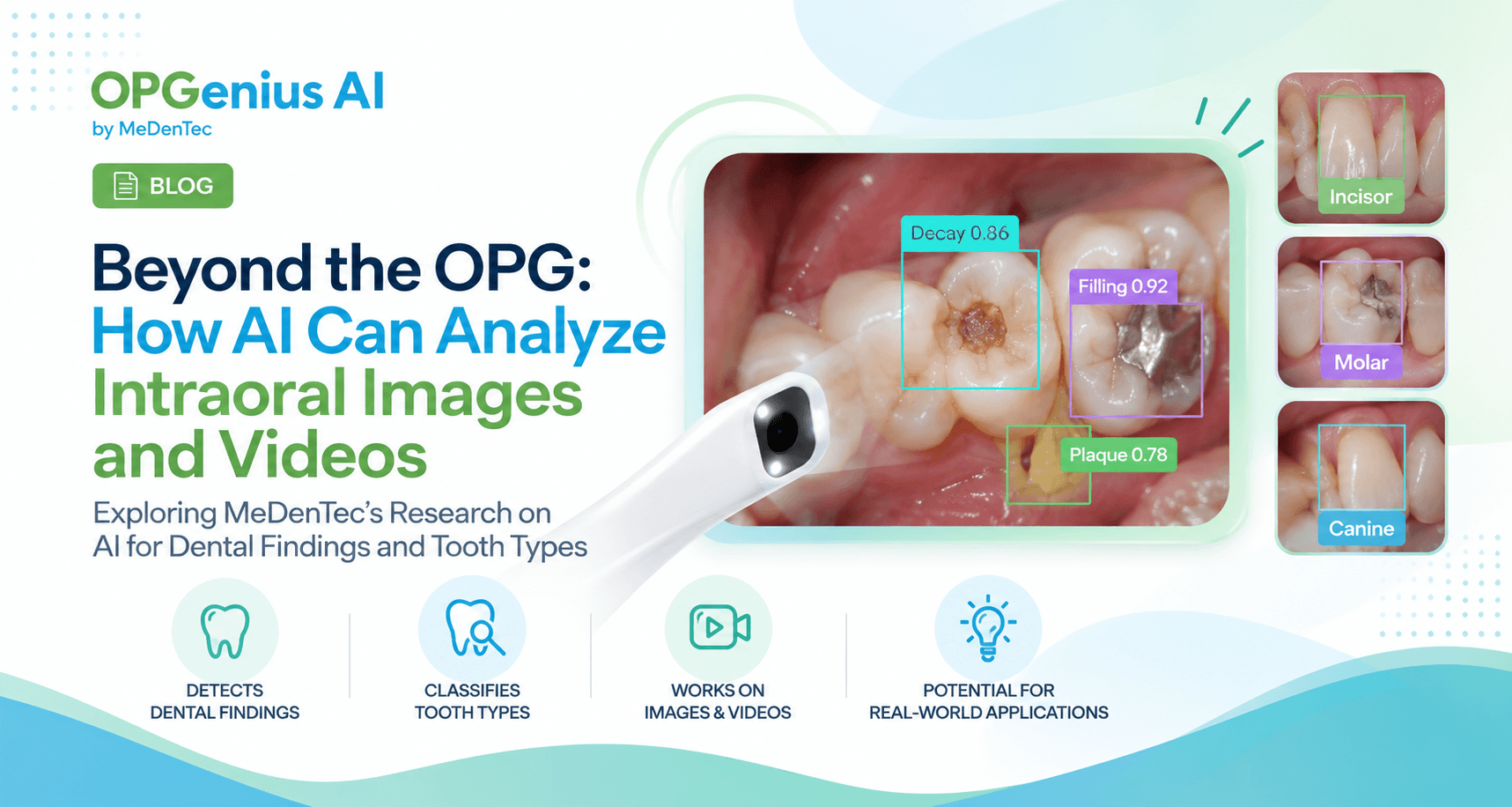
A dentist might recognize a suspicious area, an existing filling, surface staining, or visible plaque. A computer vision model can be trained to identify visual patterns associated with selected dental findings.
In our research, MeDenTec explored AI models for detecting:
Dental decay
Fillings
Plaque and calculus
Staining
Missing teeth
Tooth types: classification of teeth such as incisors, canines, premolars, and molars
The model's ability to recognize a finding depends on the image, the quality of the annotations, the training data, and how well the model performs on new examples.
An AI-detected finding is not automatically a confirmed diagnosis. Intraoral images show visible surfaces, and some dental conditions require clinical examination or radiographic assessment.
Our research investigated whether deep learning models could detect dental findings and classify tooth types using video captured through an electronic toothbrush equipped with a miniature intraoral camera1.
Rather than relying on a single still image, the project explored how frames extracted from video could provide the visual data needed to train computer vision models.
The research involved:
708 videos captured using the camera-equipped toothbrush
16,552 frames extracted from those videos
7,963 eligible frames for the dental-findings model
3,799 eligible frames for the tooth-type model
Dental experts annotated the relevant images to provide reference labels for model development and evaluation.
The team then trained YOLOv8-based models to identify selected dental findings and classify tooth types.
The research represents a proof of concept for applying computer vision to dental video captured through an everyday oral-care device.
So, how well did the models perform?
The published study reported the following results:
Dental-findings model
83.0% mAP
F1 score: 77.9%
Tooth-type model
88.6% mAP
F1 score: 86.2%
The study also reported a 94.5% mAP for amalgam-filling detection and an 84.2% mAP for decay detection.
These results demonstrate that AI models can learn to identify selected dental patterns in toothbrush-captured video frames.
But there's an important distinction between promising model performance and proven clinical effectiveness.
Further validation is needed to establish how such systems perform across different people, oral conditions, camera configurations, and real-world clinical settings.
But why analyze video instead of only still images?
Here's an interesting question: if a single image can contain useful information, why use video?
Video offers a sequence of frames rather than one isolated view. As a camera moves around the mouth, it may capture different teeth, surfaces, and viewing angles.
That creates opportunities for AI-assisted analysis, but also introduces challenges.
For example:
Some frames may be blurry.
Lighting may change as the camera moves.
The same tooth may appear in multiple frames.
Some surfaces may never be captured clearly.
Motion can make small details difficult to interpret.
A model must learn to recognize relevant patterns despite these variations.
And detecting a feature in an individual frame is not the same as reliably assessing the whole mouth.
More frames can provide more visual information, but they don't guarantee a complete or accurate assessment.
And where could this technology be useful?
The long-term potential extends beyond the research setting.
Remote oral-health support
Intraoral images could potentially help dental professionals review visible findings remotely, provided the images are suitable and the workflow is appropriately validated.
Patient education
Visual AI markers may help people understand what a dental professional is drawing attention to and prepare questions for a consultation.
Oral-health monitoring
Repeated images could potentially support observation of visible changes over time. This would require consistent image capture and validation of the monitoring approach.
Community and preventive care
In settings where access to dental professionals is limited, intraoral imaging combined with AI could be investigated as a tool to support screening and referral workflows.
However, these are potential applications,not proof that the research model is already suitable for independent screening, diagnosis, or clinical decision-making.
The next stage of research involves more than improving model metrics.
Researchers need to understand how models perform on independent datasets, how image quality affects their predictions, which findings are difficult to detect, and whether the technology provides meaningful benefits in real-world workflows.
It's also important to consider privacy, informed consent, data quality, and how AI-generated results are communicated to users.
At MeDenTec, our research into intraoral video is part of a broader exploration of how computer vision can be applied to dental imaging and oral healthcare.
Because dental AI isn't limited to one type of image or one way of collecting data.
Different imaging methods create different opportunities and different challenges.
An OPG offers a broad view of the teeth and jaws. Intraoral imaging offers a closer look at visible tooth surfaces.
Interested in how dental AI models are built, trained, and evaluated?
Explore more from MeDenTec
Want to see AI-powered dental analysis in action?
Try a Free OPG Scan